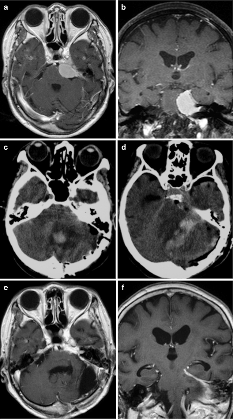
Fig. 3.
Representative patient (case 5) with life-threatening venous complications. Preoperative contrast-enhanced axial (a) and coronal (b) magnetic resonance (MR) images revealed a homogeneous mass at the left cerebellopontine angle with attachment to the tentorium. Postoperative computed tomography scans obtained at one day after the first operation (c and d) demonstrated intracerebellar hemorrhage and swelling. Postoperative contrast-enhanced axial (e) and coronal (f) MR images obtained at 3 months after the operation showed total tumor resection and atrophic cerebellum

