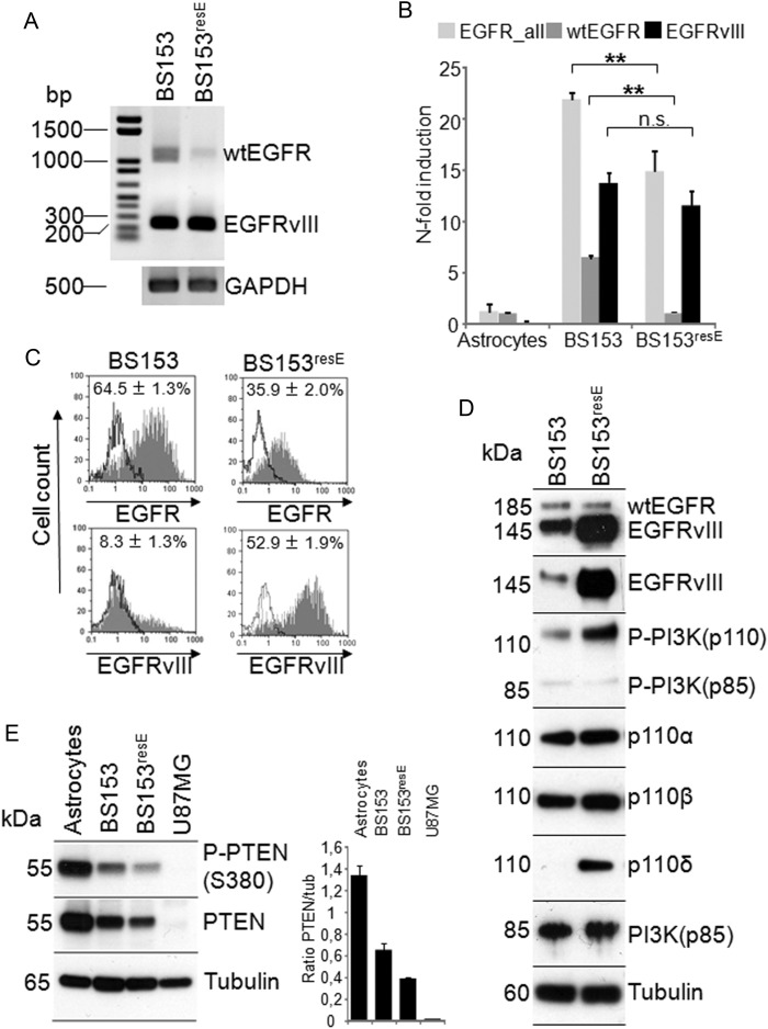
Fig. 4.
Comparison of BS153 and BS153resE at the mRNA and protein level. cDNA was analyzed by conventional PCR for the expression of EGFRvIII and wtEGFR mRNA (A) showing strong EGFRvIII expression in both cell lines and reduced wtEGFR expression in BS153resE. Quantitative real-time PCR confirmed reduced expression of wtEGFR mRNA in BS153resE compared with BS153 (**P < .005, t-test), while EGFRvIII mRNA remained unchanged. GAPDH, glyceraldehyde 3-phosphate dehydrogenase. Astrocyte cDNA was used as calibrator (B; values are means ± SD of quadruplicate determinations). Strongly enhanced expression of EGFRvIII protein and decreased expression of wtEGFR protein using variant-specific antibodies was detected in BS153resE by flow cytometry (C). Western blot analysis of overall EGFR levels using a pan–EGFR antibody as well as selective detection of EGFRvIII using a specific antibody confirmed increased expression of EGFRvIII at the protein level. The western blot for the different PI3K isoforms showed increased PI3Kp110 phosphorylation along with increased expression of PI3Kp110δ in BS153resE (D). PTEN protein levels were decreased, but PTEN was still phosphorylated in both cell lines compared with normal human astrocytes. U87MG served as a negative control (E).

