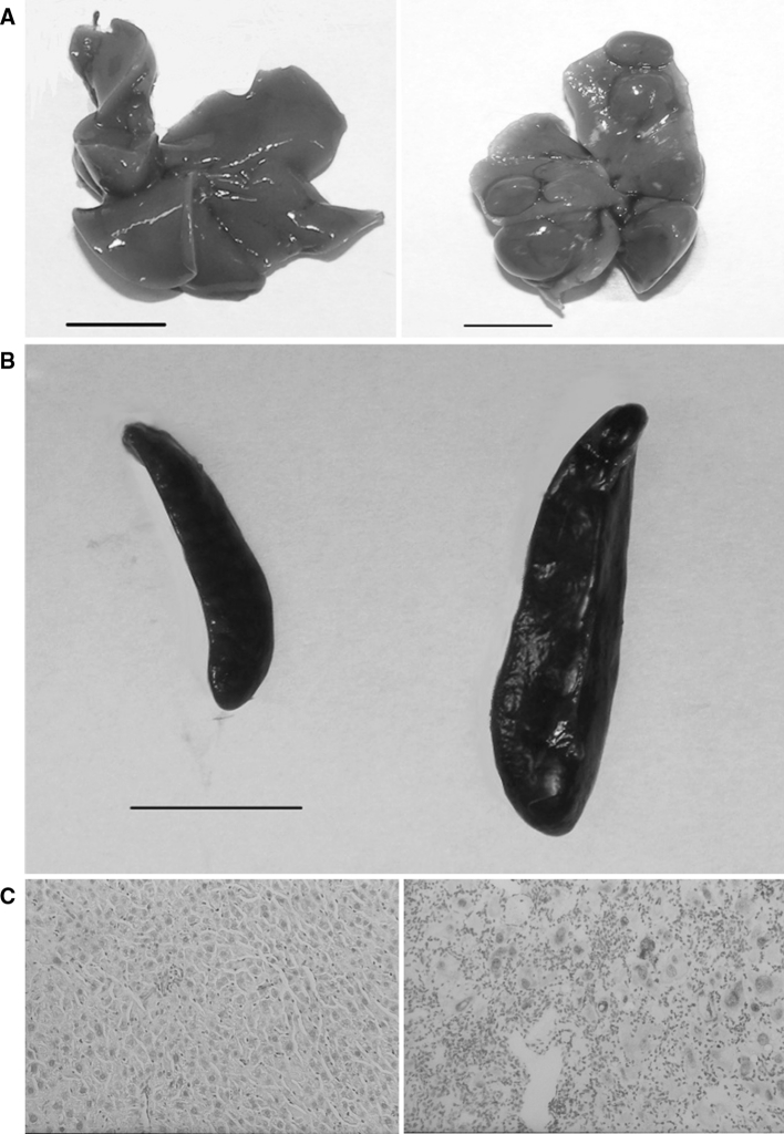
Fig. 1.
Macroscopic and microscopic changes in the organs of toxic milk mice. Control mouse liver (left) and toxic milk mouse liver (right) illustrating pathological changes at 12 months, scale bars 1 cm. a Normal spleen (left) of control mouse and enlarged spleen (right) of toxic milk mouse, scale bar 1 cm. b Unremarkable histology of the 12-month-old control mouse liver (left). Enlarged hepatocytes, inflammation, necrosis and copper deposits (red) in the liver of 12-month-old toxic milk mouse (right). Rhodanine with Mayer’s haematoxylin staining, original magnifications: ×200 (c) (Color figure online)

