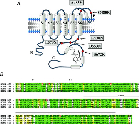
Figure 1.

A, schematic diagram of HCN topology showing one of the four channel subunits. The six transmembrane domains (S1–S6) and the intracellular N- and C-termini are shown. The C-terminus incorporates the cyclic nucleotide binding domain (CNBD) and a bound cAMP molecule. Also shown are approximate positions of HCN4 mutations reported to be linked to rhythm disorders. B, sequence alignment of the four human HCN isoforms from the pore (P) domain through to the CNBD. The position of the mutated HCN4 residues are also indicated (red background).
