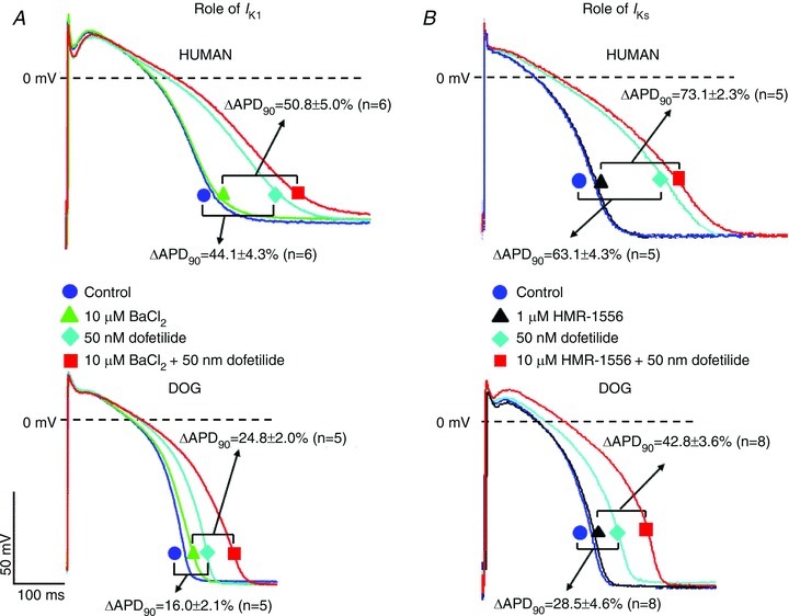
Figure 6. Effect of combined IKr+IK1 and IKr+IKs inhibition in human and dog ventricular muscle preparations (endocardial impalements).

A, representative APs at baseline (circle), following exposure to 10 μmol l−1 BaCl2 (triangle), 50 nmol l−1 dofetilide (diamond), and combined 10 μmol l−1 BaCl2+ 50 nmol l−1 dofetilide (rectangle) in human (top traces) and dog (bottom traces) ventricular muscle. Brackets show average differences between conditions indicated. B, representative APs at baseline (circle), following exposure to 1 μmol l−1 HMR-1566 (triangle), 50 nmol l−1 dofetilide (diamond), and combined 1 μmol l−1 HMR-1566 + 50 nmol l−1 dofetilide (rectangle) in human (top traces) and dog (bottom traces) ventricular muscle. Brackets show average differences between conditions indicated.
