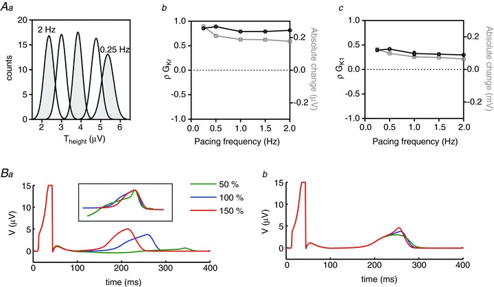
Figure 8. Rate dependence of Theight sensitivities.

Aa, Theight distributions at pacing rates between 0.25 and 2 Hz. Ab and c, rate dependence of PLS regression coefficients (ρ, black line) and absolute changes (grey line) in Theight associated with GKr (b) and GK1 (c). B, pseudo ECGs simulated with baseline and ±50%GKr (a) and GK1 (b). The inset in Ba shows T waves normalised to their peaks illustrating that GKr variability primarily affects the early part of the T wave.
