
Figure 1. Model construction.
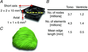
A, axial torso scans and short-axis cine scans of the paediatric CHD patient. B, the number of nodes, number of elements and mean edge length of the resulting torso mesh, and of the ventricular portion of the mesh. C, streamlined image of the fibre orientations in the ventricular mesh.