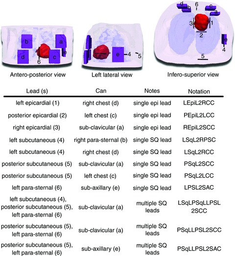
Figure 2. Implantable cardioverter–defibrillator placement.

A, the finite element heart–torso mesh and ICD can (purple; a–e) and ICD lead (black; 1–6) placement locations. The ventricles are shown in red, skin in transparent pink, bones in transparent white and lungs in transparent blue. Segmented and modelled, but not shown here, were fat, blood, muscles and remaining conductive medium. B, the 11 ICD configurations that were tested in this study (characters and numbers in parentheses refer to A). Three configurations used epicardial (epi) leads, five used single subcutaneous (SQ) leads and three used multiple subcutaneous leads.
