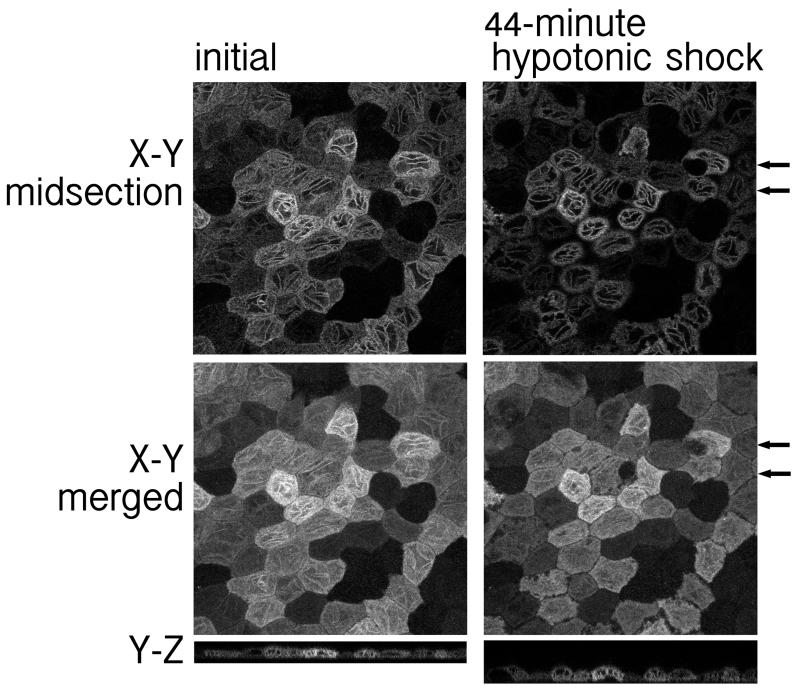
FIGURE 6.
The linear fluorescent tubulin-like structure (TLS) found in well-differentiated MDCK cells expressing SLC13A3_15 persisted after a 44-minute hypotonic shock. (Left) Images of XY midsection and merged XY and YZ sections in Hanks’ balanced salt solution with 0.5% FBS. (Right) Images of XY midsection and merged XY and YZ sections of cells treated with 25% osmolarity hypotonic shock at 37° C incubation for 44-minute. Hypotonic condition was created by adding 1.5 ml sterile water to the 0.5 ml Hanks’ balanced salt solution with 0.5% FBS in the chamber. Arrows: the location of cells that became deformed during the shock.

