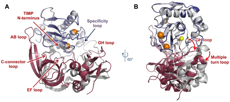
Figure 4. Comparison of MMP-10cd/TIMP-1 and MMP-10cd/TIMP-2 complexes.
MMP-10cd/TIMP-2 molecules are shown in blue and raspberry, respectively, with MMP-10cd/TIMP-1 complex (PDB ID: 3V96) [24] shown in white; complexes are superposed based on Cα atoms of all MMP-10cd residues. (A) The long AB loop of TIMP-2 forms a much more extensive contact area with the MMP-10cd than is seen with TIMP-1, while the C-terminal loops of TIMP-2 form fewer contacts than in the complex with TIMP-1. (B) TIMP-2 is rotated by ∼21° around an axis centered on the catalytic zinc when compared with TIMP-1.

