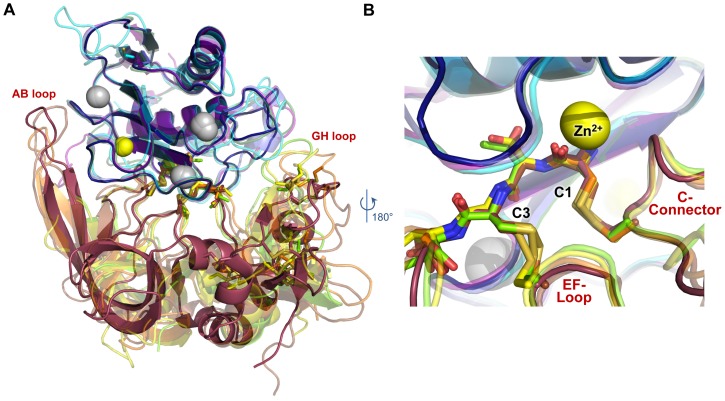
Figure 5. MMP/TIMP complexes feature conserved core interactions but highly diverse peripheral interactions.
MMP-10cd/TIMP-2 (indigo/raspberry) is superposed with four different MMPcd/TIMP structures based on Cα atoms of all corresponding MMP residues: MMP-10cd/TIMP-1 (slate/chartreuse; PDB ID: 3V96) [24], MMP-3cd/TIMP-1 (purple/yellow; PDB ID: 1UEA) [26], MT1-MMPcd/TIMP-2 (cyan/brown; PDB ID: 1BQQ) [27], and MMP-13cd/TIMP-2 (forest/orange; PDB ID: 2E2D) [28]. (A) Positioning of peripheral TIMP loops including the AB and GH loops relative to the MMP show wide variability. (B) In the MMP active site, backbone positioning of TIMP residues 1–4 and the C-connector loop are nearly identical.

