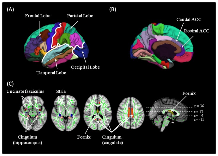
Figure 1. Grey matter and white matter regions of interest used in the study.
(top row) Different brain lobes (A) and the rostral and caudal anterior cingulate sub-regions (B) parcelled using freesurfer for a representative dataset (bottom row (C)). Fractional anisotropy skeleton (in green) representing the major white matter tracts for all subjects is overlaid on the MNI standard brain. Sections of the tract sekeleton representing the cingulate and limbic white matter tracts are shown: cingulum cingulate bundle (orange); cingulum hippocampus bundle (pink); fornix (yellow); stria (blue) and uncinate fasciculus (red).
Abbreviations: ACC, anterior cingulate cortex.

