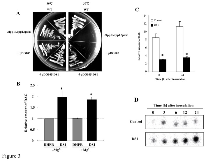
Figure 3. Functional analysis of DS1.
(A) Isogenic yeast strain ∆lpp1∆dpp1∆pah1 containing empty pDO105 plasmid (+pDO105) or pDO105 containing DS1 (+pDO105: DS1) were cultured on agar YPD plates and incubated at 30°C or 37°C. (B) PAP activity of DS1 and dehydrofolate reductase (DHFR; negative control) was determined in the presence or absence of Mg2+ as described in Materials and Methods. Values are means and SD from triplicate experiments. Asterisks denote values significantly different from those of control (*; P < 0.05). (C) PAP activity in control (white bar) and DS1 plants (black bar). Crude protein fractions were isolated from control and DS1 plants 0 and 24 h after inoculation with R. solanacearum. PAP activity was determined without Mg2+ as described in Materials and Methods. Values are means and SD from triplicate experiments. Asterisks denote values significantly different from those of control (*; P < 0.05). (D) Phosphatidic acid contents in control and DS1 plants. Total lipid fraction was extracted from control and DS1 plants 0 to 24 h after inoculation with R. solanacearum. PA was separated by ethyl acetate TLC as described in Materials and Methods.

