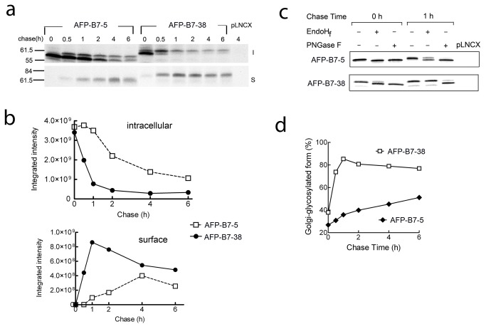
Figure 4. AFP intracellular transport can be followed by AFP glycosylation status.
a) 3T3 cells stably expressing AFP-B7-5 or AFP-B7-38 were pulsed with 35S-methionine and chased with cold methionine for the indicated times. Surface (S, lower panel) or intracellular (I, upper panel) AFP at each time is shown. Vector-transfected control 3T3 cells are indicated as pLNCX. b) Protein bands in panel a were quantified to show the temporal change in 35S-methionine labeled intracellular (upper panel, both upper and lower bands) and surface (lower panel) AFP chimeric proteins. c) 3T3 cells stably expressing AFP-B7-5 or AFP-B7-38 were pulsed with 35S-methionine and then chased with cold methionine for 0 or 1 h. AFP immunoprecipitates were untreated or treated with EndoHf or PNGaseF deglycosylase as indicated. d) Quantification of the percentage of intracellular AFP chimera with complex-type N-linked carbohydrate (Golgi-glycosylated form) in relation to total intracellular AFP chimeric protein in 35S-methionine labeled cells at the indicated chase times.

