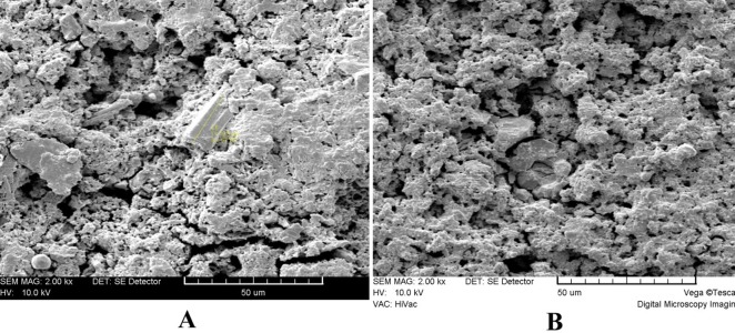
Figure 4 .

Surface microstructure of Blood-exposed MTA (×2000). (A) Numerous microchannels and few rounded hexagonal crystals partially covered by a gel form structure are evident. No needle like structures can be seen. (B) The crystals tend to be more rounded and partially covered by a poorly-crystalline structure.
