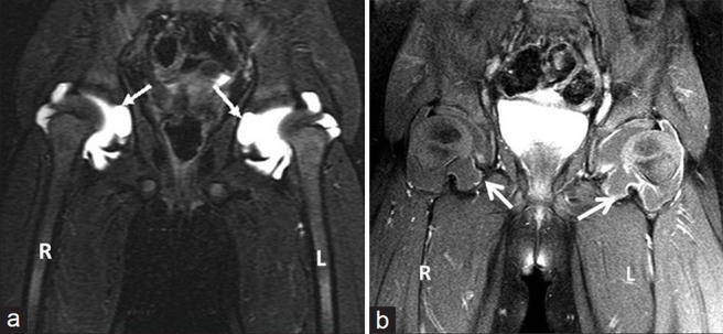
Figure 3.

(a) Three-year 3-month-old girl child with CACP syndrome. Coronal STIR image of hips (on 3T system) shows large bilateral joint effusions. Note the intra-osseous acetabular herniations communicating with the effusions (arrows) and (b) 3-year 3-month-old girl child with CACP syndrome. Coronal T1-weighted image of hips (on 3T system) obtained after administration of gadolinium shows rim enhancement of joint capsule (better appreciated on the left side) and of walls of intraosseous herniations (arrows). Note the lack of thick solid synovial enhancement as seen in Juvenile idiopathic arthritis.
