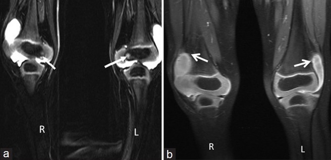
Figure 4.

(a) Three-year 3-month-old girl child with CACP syndrome. Coronal STIR image of both knees (on 3T system) shows large effusions. Small intraosseous cysts are identified along the medial femoral condyle (arrows) and (b) 3 year 3 month old girl child with CACP syndrome. Coronal T1 weighted of both knees (on 3T system) shows effusion with mildly thickened synovium (arrows).
