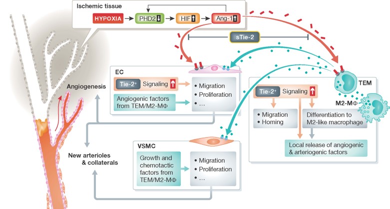
Figure 1. Molecular and cellular mechanisms initiating by Angiotensin-1 and leading to post-ischemic vascular repair.
In ischemic tissues, the activity of the oxygen sensor PDH2 is reduced, permitting the HIFα-mediated transcription of pro-angiogenic genes, such as Ang-1 (red). In a feedback loop, Ang-1 after binding with its Tie2 receptor, represses PHD2. This leads to increased circulating and ischemic tissue-engrafted TEM and skews macrophages to the proangiogenic M2 type. TEM and M2 release pro-angiogenic/pro-arteriogenic factors (blue) acting on endothelial cells (EC) and vascular smooth muscle cells (VSMC) to promote cellular processes leading to formation of capillaries, arterioles and arterial collaterals. Additionally, Ang-1 and Ang-2 binding to Tie2 receptor regulates angiogenesis at EC level. Soluble Tie2 (sTie2) inhibits Ang/Tie2 signalling in ischemia, thus contrasting vascular repair.

