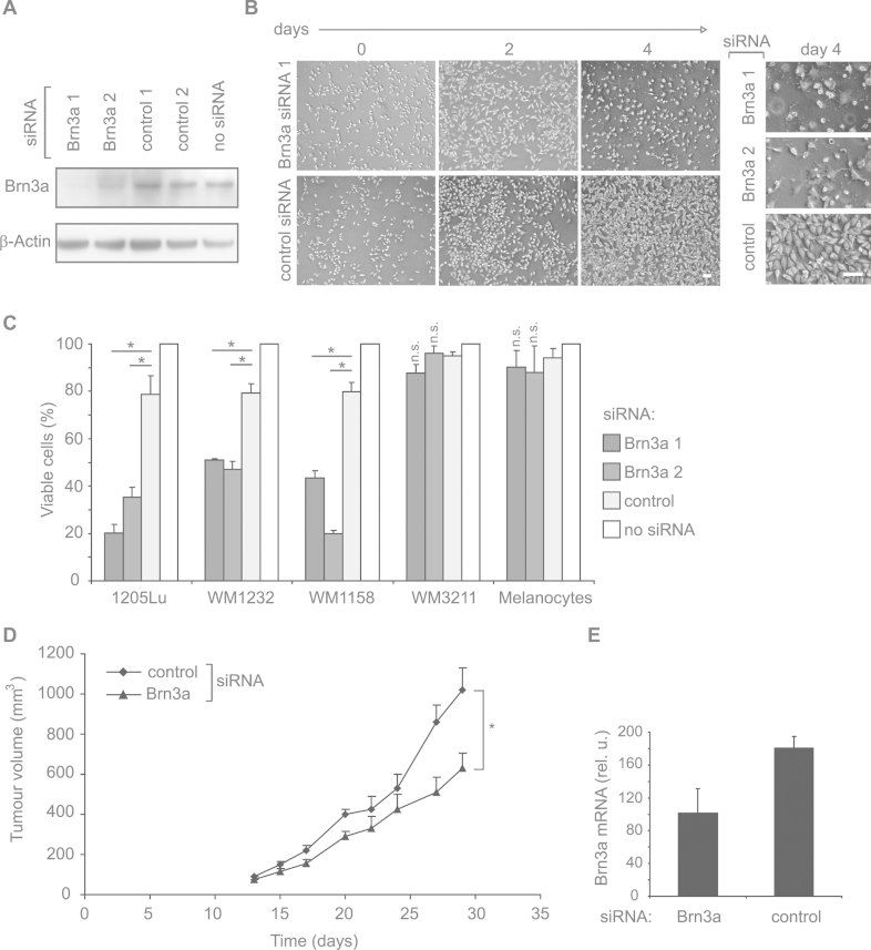
Figure 2. Inhibition of Brn3a reduces melanoma cell viability and leads to reduced tumour growth in vivo.
- 1205Lu melanoma cells were transfected with two siRNAs specific for Brn3a (Brn3a 1 and Brn3a 2) or control siRNAs and analysed by immunoblotting 48 h after transfection. Representative blots (n = 3) are shown.
- Microscopic pictures of 1205Lu melanoma cells 2 or 4 days after transfection of Brn3a-specific siRNAs or control siRNA. Scale bars: 100 µm.
- Different melanoma cell lines, i.e. 1205Lu, WM1158 and WM1232 (high Brn3a levels) and WM3211 (low Brn3a levels), were transfected with siRNAs as described in (A). Viable melanoma cells were quantified 4 days after siRNA transfection. Viability of cells treated with transfection reagent alone (‘no siRNA’) was set to 100%. Mean ± SD is shown. *p = 0.003 or less, t-test, n = 3 per group; n.s.: not significant (p > 0.05).
- 1205Lu melanoma cells were subcutaneously injected into nude mice. Upon palpability of tumours, mice were systemically treated three times per week by intraperitoneal injection of 10 µg of Brn3a-specific or control siRNA complexed with polyethylenimine. Tumour growth in Brn3a siRNA-treated animals was significantly reduced (p = 0.0078, Wilcoxon matched pair test, n = 5 per group).
- Brn3a mRNA levels in tumours isolated at day 29. Mean of each group ± SEM is shown.

