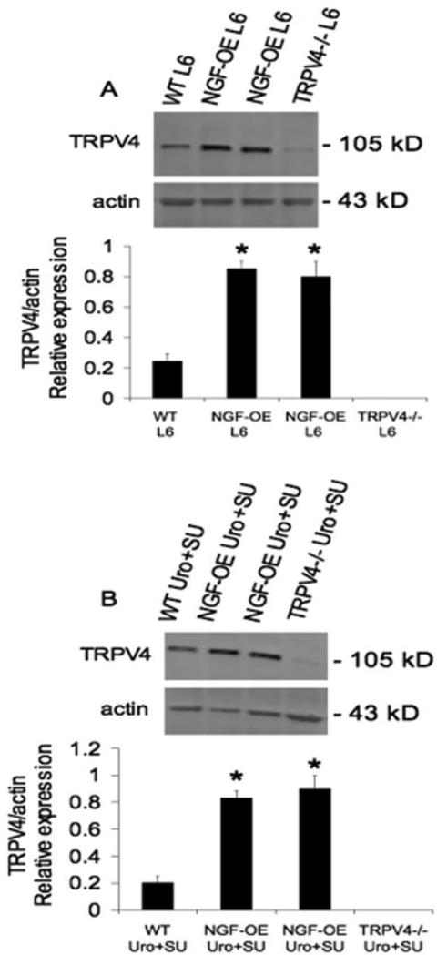
Figure 3.
Western blot analyses of TRPV4 expression in lumbar (L) 6 dorsal root ganglia (DRG) (A) and urothelium (Uro) + suburothelium (SU) from wildtype (WT), NGF-overexpressing (OE) and TRPV4-/- mice. Representative western blot of TRPV4 protein expression in L6 DRG from WT, NGF-OE and TRPV4-/- mice (A). Summary histogram of the relative expression of TRPV4 protein normalized to actin expression in L6 DRG from WT, NGF-OE and TRPV4-/- mice (A). Representative western blot of TRPV4 protein expression in Uro + SU from WT, NGF-OE and TRPV4-/- mice (B). Summary histogram of the relative expression of TRPV4 protein normalized to actin expression in Uro + SU from WT, NGF-OE and TRPV4-/- mice (B). Samples size are n of 7 – 9; *, p ≤ 0.001 versus WT.

