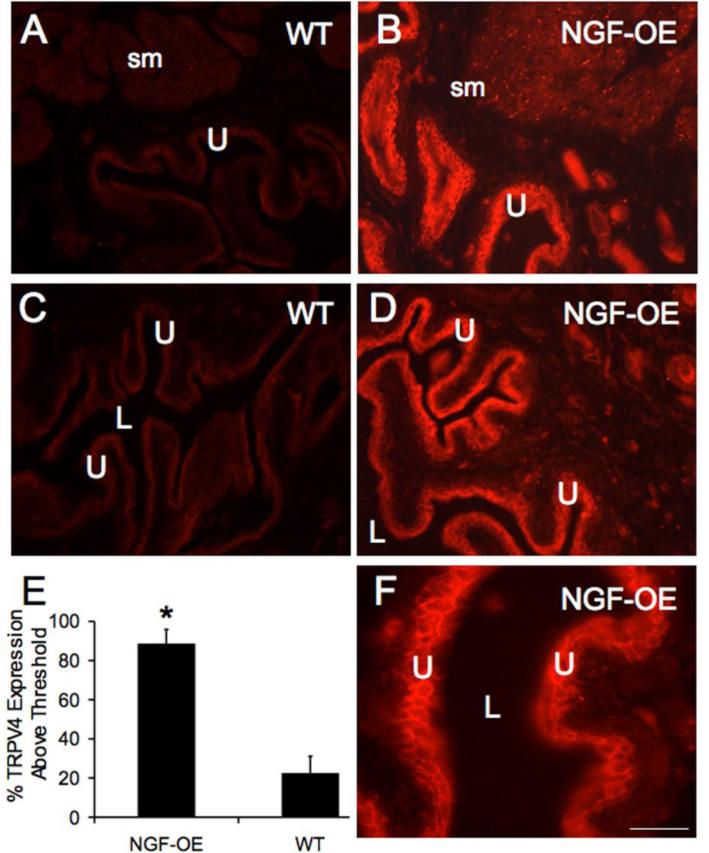
Figure 4.
TRPV4-immunoreactivity (IR) in cryostat sections of urinary bladder from WT and NGF-OE mice. In WT mice, faint TRPV4-IR was present in urothelium (U) and detrusor smooth muscle (sm) (A, C). Chronic overexpression of NGF in the U increased TRPV4-IR in U (B, D, F) and detrusor sm (B). Higher power fluorescence image of TRPV4-IR in U (F). TRPV4-IR was exhibited in all cellular layers of the U (A-D, F) in WT and NGF-OE mice. TRPV4-IR was present in the U throughout the bladder dome, body and neck regions. For all images, exposure times were held constant, and all tissues were processed simultaneously. E: Histogram of TRPV4 expression above threshold in the U of NGF-OE and WT mice expressed as a percentage of control. E. TRPV4-IR above threshold in U was significantly (p ≤ 0.001) increased in NGF-OE mice. Calibration bar represents 50 μm in A-D and 25 μm in F. Data are a summary of n = 7-9 for each group. L, lumen.

