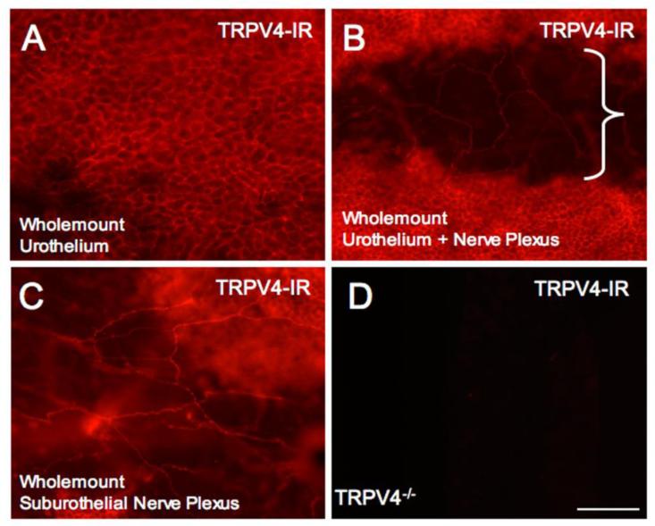
Figure 6.
TRPV4-immunoreactivity (IR) in wholemount preparations of the urinary bladder from NGF-OE mice. In wholemount preparations, robust TRPV4-IR was present throughout the urothelium in the bladder dome, body and neck regions (A, B). With the urothelium dissected, TRPV4-IR is observed in the suburothelial nerve plexus (B, bracketed region). The bracketed region (B) is an area with the urothelium removed showing the suburothelial nerve plexus as in C. TRPV4-IR was present in the suburothelial plexus (C) in the bladder dome, body and neck and with greatest density of TRPV4-immunoreactive nerve fibers being present in the bladder neck. The specificity of the TRPV4 antibody used was verified in urinary bladder harvested from TRPV4-/- mice that did not exhibit TRPV4-IR (D). Calibration bar represents 50 μm in A - D.

