
Figure 7. The aged-associated increase in brain levels of miR-29a and miR-29b was negatively correlated with reduced expression of CX3CL1 and IGF-1.
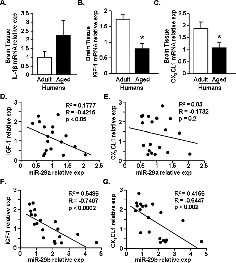
Postmortem brain tissue was acquired for both adult (n=9) and aged (n=13) age groups. From the human brain tissue mRNA expression of A) IL-1β, B) IGF-1, and C) CX3CL1 was determined. In addition, a correlation plot was created for IGF-1 and CX3CL1 compared to miR-29a or miR-29b levels determined in Fig.5. Plots depict expression levels of miR-29a versus D) IGF-1 and E) CX3CL1 in the human brain tissue. In addition, levels of miR-29b versus F) IGF-1 and G) CX3CL1 are shown. Bars represent the mean ± SEM. Relative expression is compared to the average comparative Ct for all samples. Means with * are significantly (p<0.04) different from Adult controls.