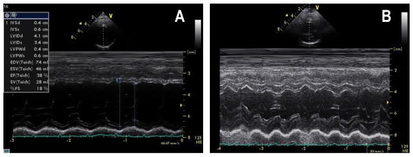
Figure 1.

2D guided left ventricular M-mode echocardiography showing in case 1 at 4y normal left ventricular function and left ventricular fractional shortening of 35%(B), and case 2 at 3y with dilated left ventricle, reduced left ventricular function with fractional shortening of 18% (A). S=systole, D=diastole, LV=left ventricle, RV-right ventricle, LVPW=left ventricle posterior wall, IVS=inter-ventricular septum, FS=fractional shortening
