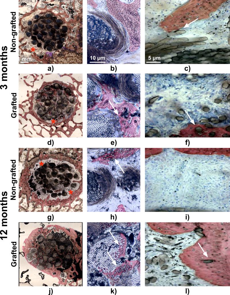
Fig. 8.
Light micrographs of representative histological sections. a-c) and d-f) non-grafted and grafted ligament respectively at 3 months post implantation, g-i) and j-l) non grafted and grafted ligaments respectively at 12 months post-implantation. Red stars indicate the fibrovascular connective tissue at the bone-ligament interface and white arrows show the PET fibres embedded in the newly formed bone. Stevenels’ blue (cell nuclei) and van Gieson picro-fuschin (bone tissue).

