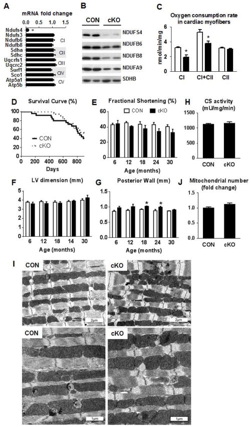
Figure 1. Mouse survival, cardiac function and mitochondrial assessment in Ndufs4 deficient mice.
(A) Fold changes ±SEM of ETC gene expression in the cKO mice relative to CON (n=7). (B) Representative western blot for ETC proteins (n=4). (C) Mitochondrial state 3 respiration in permeabilized cardiac fibers. Pyruvate + malate was used as complex I substrate in the presence of ADP, then succinate was added to establish complex I + complex II (CI+CII) respiration and finally rotenone was added to block complex I and establish complex II (CII) respiration (mean ±SEM; n=3). (D) Kaplan-Meier survival curve of cKO and CON mice (n=19–25). Echocardiographic data depicting (E) fractional shortening (%), (F) LV end-diastolic dimension (mm) and (G) posterior wall thickness (mm) in CON (white) and cKO (black) mice over 30 months (n=6–13). (H) Cardiac tissue citrate synthase enzyme activity (n=13–15). Data are expressed as means ± SEM. *P<0.05 vs CON. (I) Electron microscopy images illustrating mitochondrial arrangement and morphological characteristic and (J) fold differences in total mitochondrial number. *P<0.05 vs CON. See also Figure S1 and Table S1.

