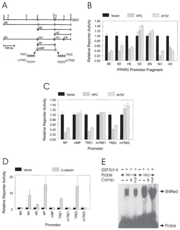
Figure 2. APC Regulates PPARδ Expression through CRT.
(A) PPARδ promotor. A restriction map of the 3.1 kb region upstream of the first exon of PPARδ is shown. Restriction fragments BE, NE, HE, DE, BN, NH, HD, and NP were used to construct reporters for measuring APC and β-catenin responsiveness. Filled boxes represent potential Tcf-4-binding sites, and open boxes represent the same sites engineered to contain mutations that abolish Tcf-4 binding. mNP represents fragment NP with both potential Tcf-4-binding sites mutated. TRE1 and TRE2 contain four repeats of the two Tcf-4-binding sites, respectively. mTRE1 and mTRE2 are mutant forms of TRE1 and TRE2.
(B) The PPARδ promotor is repressed by APC and dnTcf. SW480 CRC cells were transfected with the indicated PPARδ promotor luciferase reporters (0.4 μg), with a β-galactosidase expression vector (0.2 μg pCMVβ), and with 1.0 μg of either a control (Vector), APC, or dnTcf expression vector. Luciferase activity is reported relative to the vector control after normalizing for transfection efficiency through β-galactosidase activity. Bars represent the means of three independent replicates with error bars representing the unbiased standard deviations.
(C) APC and dnTcf responsiveness is mediated by two putative Tcf-4-binding sites. PPARδ promotor fragments with intact and mutated Tcf-4-binding sites were tested for APC and dnTcf-4 responsiveness as described in (B). Bars represent the means of three independent replicates, with error bars representing the unbiased standard deviations.
(D) β-catenin transactivation maps to the same promotor regions mediating APC and dnTcf responsiveness. 293 cells were transfected with the indicated PPARδ promotor luciferase reporters (0.4 μg), with a β-galactosidase expression vector (0.2 μg pCMVβ), and with 1.0 μg of either a no insert control (Vector) or an oncogenic β-catenin (β-catenin) expression vector. Luciferase activity was reported as described for (B). Bars represent the means of three independent replicates, with error bars representing the unbiased standard deviations.
(E) Putative Tcf-4-binding sites in the PPARδ promotor bind Tcf-4. GEMSA was performed using 32P-labeled probes containing either putative Tcf-4-binding sites TRE1 or TRE2. GEMSA was performed in the presence of a GST fusion protein containing the Tcf-4 DNA-binding domain as indicated. Wild-type (wt) or mutant (mut) competitors corresponding to the Tcf-4-binding sites were used as indicated.

