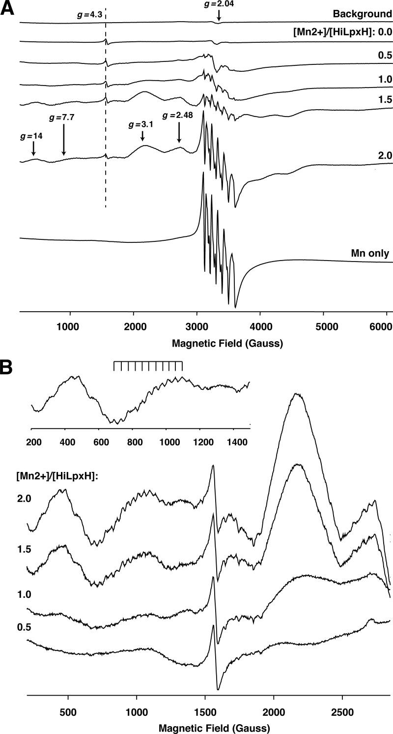
FIGURE 7.
Cryogenic EPR spectra of HiLpxH in the presence of Mn2+. A, representative continuous wave X-band EPR spectra measured at 28 K. From top to bottom are background signal from an EPR resonator outfitted with a low temperature quartz insert; signal from samples a range of [Mn2+]/[HiLpxH] ratios; and signal from a Mn2+ only solution. Position of the g = 4.3 marker originating from a low symmetry Fe3+ ion nonspecifically bound to the protein is shown by a dashed line. The areas of the spectra indicating the presence of a di-Mn2+ cluster (g = 14, 7.7, 3.1, and 2.48) are denoted by arrows. B, spectra from A enhanced in the range 500–2,500 G to highlight the increase in hyperfine splitting at g = 7.7 for greater [Mn2+]/[HiLpxH] ratios.

