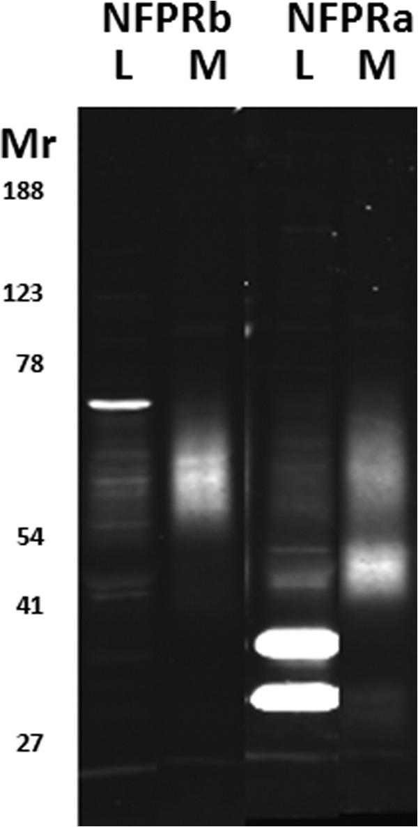
FIGURE 3.

Recognition of FPR1 and FPR2 in unstimulated neutrophil homogenates and membranes. SDS-PAGE-separated and immunoblotted whole cell lysates (L) and salt-washed membranes (M) from N2 cavitates of unstimulated neutrophils, as described under “Experimental Procedures,” were developed with the NFPRb (left two lanes) and NFPRa (right two lanes) primary mAbs and visualized by infrared fluorescence scanning with a LI-COR Odyssey scanner. Each lane contains 2 × 10⁵ cell equivalents. The broadly staining band between Mr ∼50,000 and 65,000 (also known as 60K) is putatively FPR1 and is recognized by both mAbs. The band between Mr ∼41,000 and 50,000 (also known as 45K) comigrates with FPR2 and is only recognized by NFPRa. The more intense bands at ∼30,000 and ∼36,000 are soluble cytosolic species and are lost in the particulate fraction.
