
FIGURE 2.
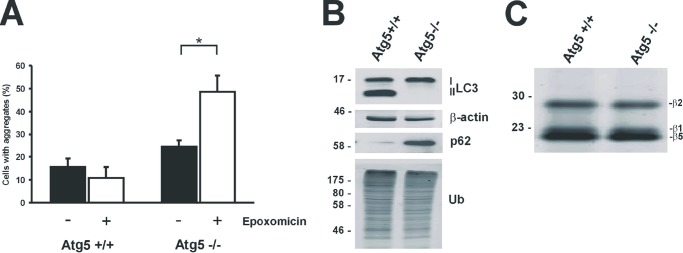
Proteasomal degradation of mHtt-exon1 in Atg5-deficient cells. A, quantification of Htt aggregates after expression of Htt-exon1-97Q-C4 in Atg5+/+ and Atg5−/− MEF cells. Six hours after electroporation, cells were treated for 16 h with DMSO or epoxomicin and fixed on coverslips for staining and aggregate scoring; *, p < 0.05 (n = 3). B, Western blot analysis for detection of endogenous LC3-I and LC3-II levels in electroporated Atg5+/+ and Atg5−/− MEF cells. C, the proteasomal catalytic sites (β1, β2, and β5) were labeled in Atg5+/+ and Atg5−/− MEF cell lysate with activity probe. Error bars, S.D.