
FIGURE 3.
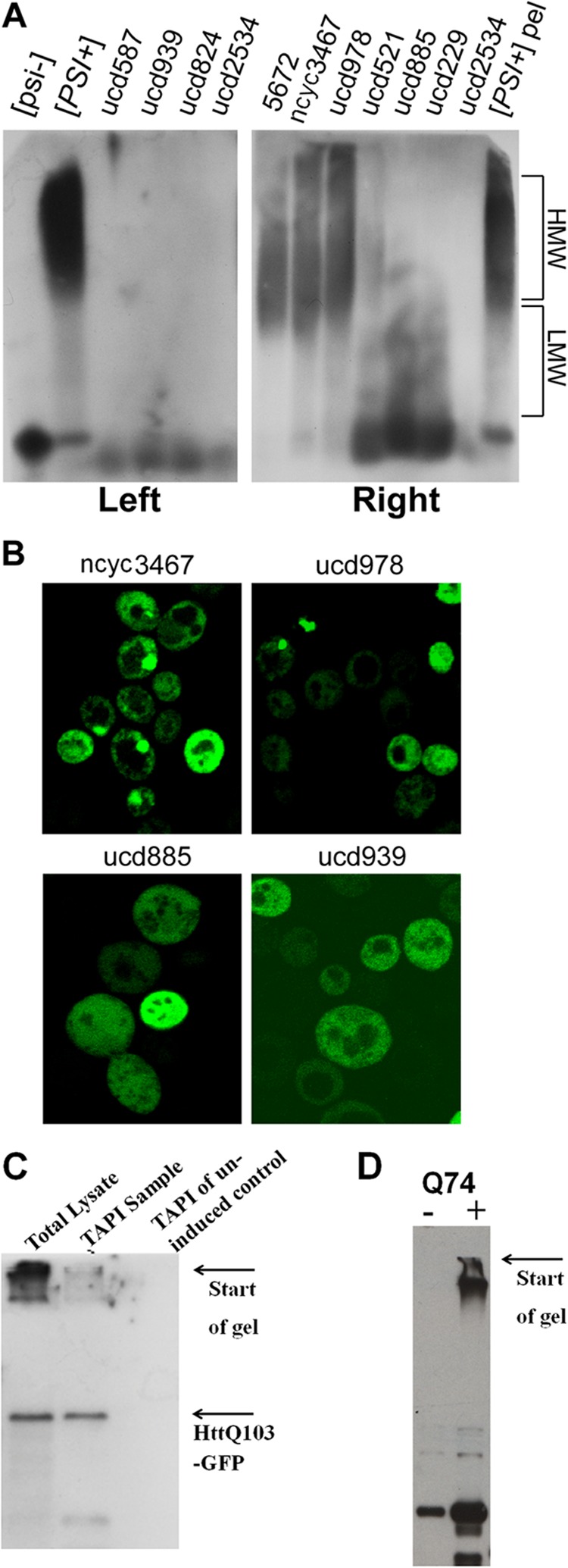
Verification of protein aggregation. A, 10 [PSI+] wild strains were analyzed by SDD-AGE (14); Sup35 was detected by immunostaining. The isogenic pair 74D-694 [PSI+][RNQ+] and [psi-][rnq-] was used as controls. HMW,- high molecular weight aggregates; LMW, low molecular weight aggregates. In the left panel, cell lysates were directly applied to SDD-AGE; in the right panel they were first pelleted (200,000 × g for 1 h), and pellets were applied to SDD-AGE (pelleting improves resolution of low molecular weight aggregates). B, the same wild strains were transformed with Sup35NM-GFP and analyzed by fluorescent microscopy. C and D, detection of polyglutamine-GFP by immunoblotting. C, yeast cells expressing HttQ103-GFP from the inducible GAL1 promoter for 5 h were lysed and subjected to TAPI. HttQ103-GFP was detected by immunoblotting with anti-GFP antibodies from crude cell lysate (lane 1) and after elution and purification before protease digestion (lane 2). No signal was detected from the uninduced control cells. D, Western analysis of the PC12 huntingtin cell model (29) with stably transfected exon 1 of the HD gene with 74 CAGs under the control of a tetracycline promoter.