
FIGURE 6.
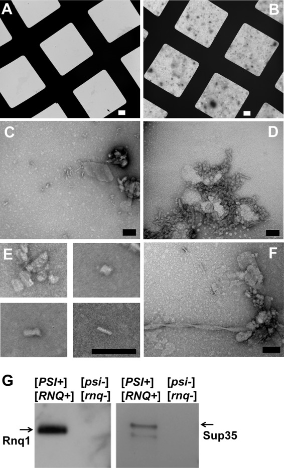
Electron micrographs of the recovered SDS-resistant protein aggregates from yeast cells. A, and B, the gel-recovered material from the isogenic strain pair 74D-694 [PSI+][RNQ+] and [psi-][rnq-] acquired at 800× direct magnification on carbon/Formvar-covered copper grids; A shows a field from the strain with no prions; B shows the field from the strain harboring both [PSI+] and [RNQ+]. C and D, representative images of the proteinaceous debris from the prion-containing strain at 80,000× and 150,000× direct magnification. E, small barrel-shaped structures observed from prion-containing samples. F, example of a fiber observed after recovered material was incubated several days at room temperature. White scale bars = 10 μm; black scale bars = 100 nm. G, Western blotting and immunostaining of the gel-recovered material from A and B above with anti-Sup35 and anti-Rnq1 antibodies.