
FIGURE 6.
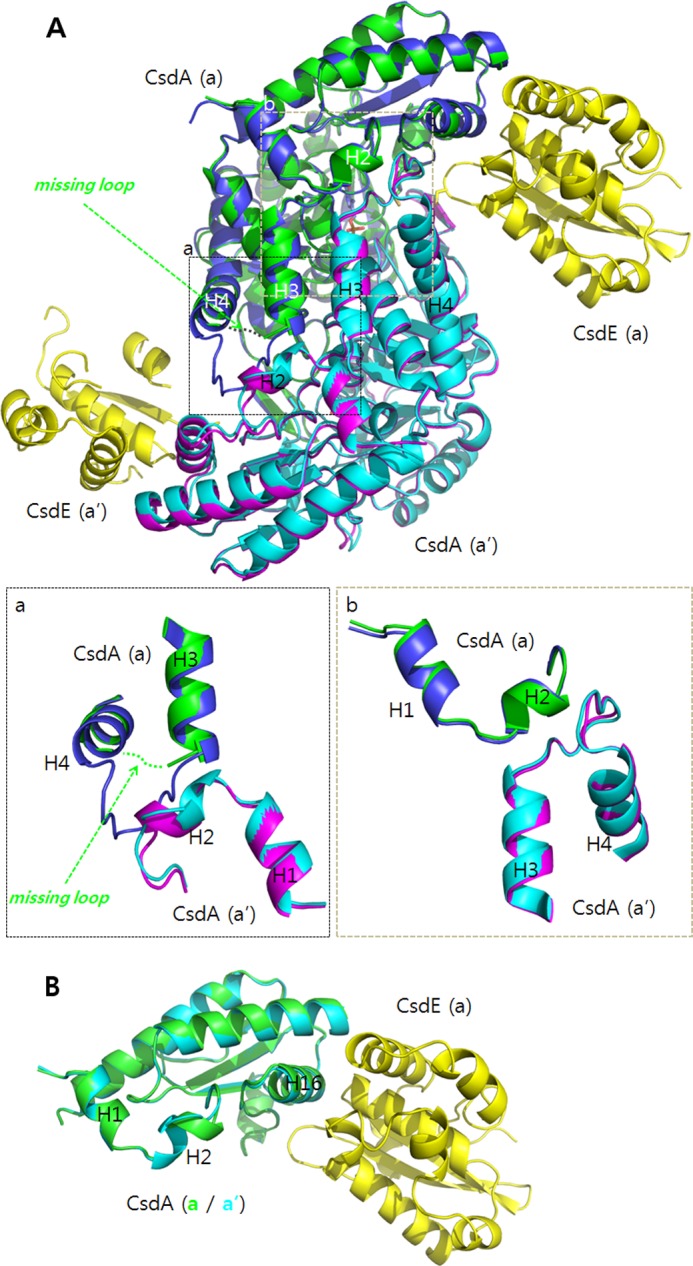
Structural differences between the free CsdA and the CsdA from CsdA-CsdE. A, two dimeric subunits of free CsdA (a in blue; a′ in violet) and CsdA from CsdA-CsdE (a in green; a′ in cyan) are structurally aligned. Both CsdEs are shown in yellow. Significant differences are seen near the dimeric interface. For one CsdA(a), part of the H3-H4 loop (CsdA residues 49–54) is disordered. The corresponding residues are ordered for CsdA(a′). The ordering in H3-H4 loop of CsdA(a′) changes the helical content of H2 in the interacting CsdA(a) (magnified in the inset, a and b). B, changes in H2 are clearly seen when the two subunits of CsdA from the CsdA-CsdE are superimposed for comparison.