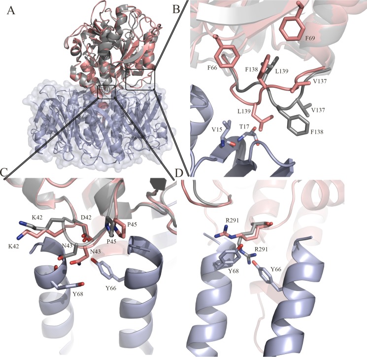
FIGURE 2.
A, overall superposition of the isolated crystal structure of SubA and the SubAB holotoxin. The crystal structure of SubA (PDB code: 2IY9) is colored in gray, and the A-subunit in the SubAB structure is colored in salmon. The B-subunit of SubAB is shown in light blue. B, structural rearrangement of the loop (Ser-135–Pro-141). C, structural rearrangement of the loop (Asp-41–Pro-45). D, structural rearrangement of the residue Arg-291 upon formation of the holotoxin. The figures were generated using PyMOL (29).

