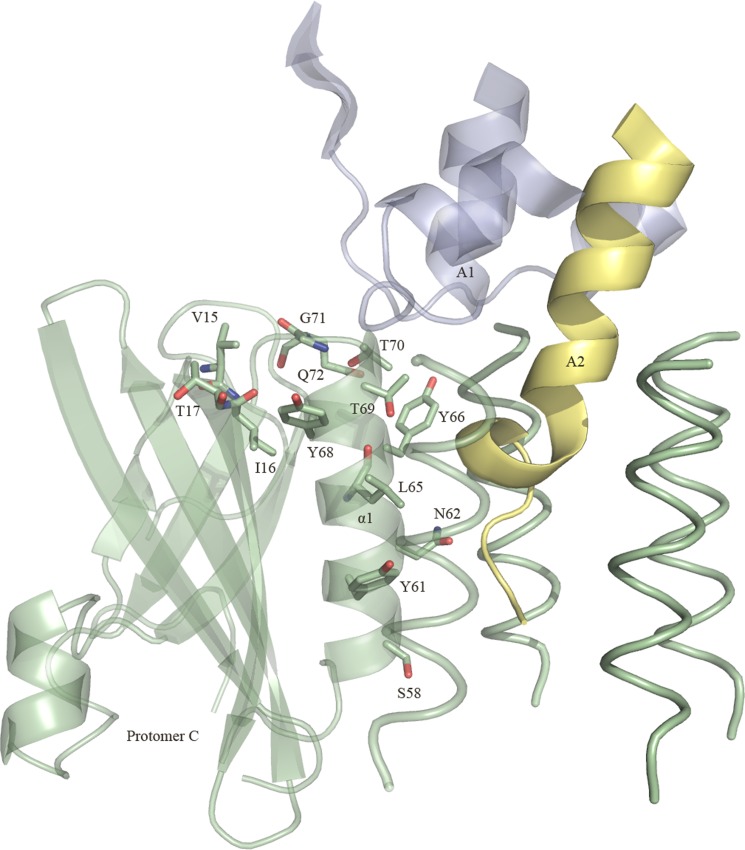
FIGURE 6.
Positions of the 13 mutated SubB residues. The locations of the residues belonging to protomer C are shown. Segments of the A1 domain and the A2 helix (SubA) are shown and colored in blue and yellow, respectively. For clarity, only the α-helix of the other four protomers is shown (in green). The figure was generated using PyMOL (29).

