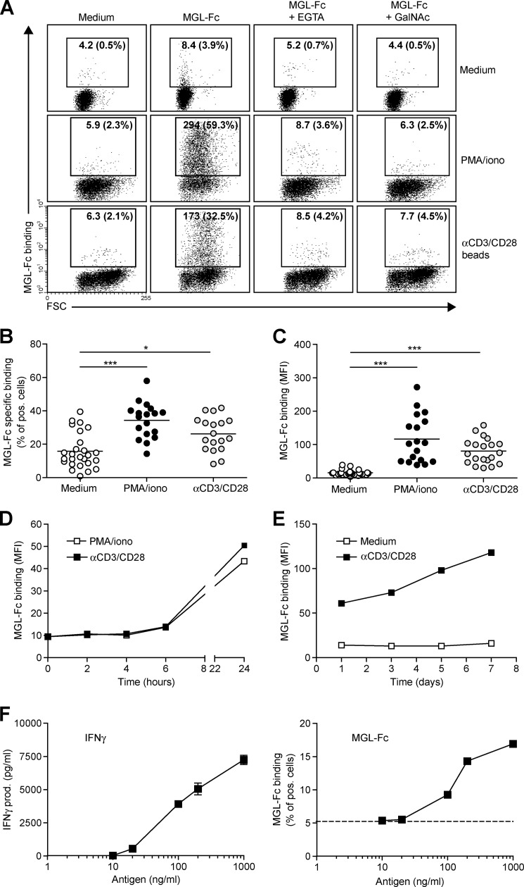
FIGURE 1.
MGL ligands are up-regulated on recently activated CD4+ T cells. CD4+ T cells were left untreated or stimulated with PMA/ionomycin or αCD3/CD28 beads. A, after overnight incubation, MGL-Fc binding in the presence or absence of EGTA or free GalNAc was measured by flow cytometry. Mean fluorescence and the % of positive cells are indicated in the plots. Data are from one representative donor out of 20. FSC, forward scatter. B and C, both the % of cells expressing MGL binding epitopes as well as the magnitude of expression increases after overnight CD4+ T cell stimulation using PMA/ionomycin or αCD3/CD28 beads. At least 18 donors are plotted for each condition. *, p < 0.05; ***, p < 0.001. D and E, MGL ligands appear after 24 h of stimulation but subsequently remain stable on the cell surface for days. CD4+ T cells were stimulated for indicated time points with medium, PMA/ionomycin, or αCD3/CD28 beads. At each time point, MGL-Fc binding was measured by flow cytometry. Experiments were repeated three times with different donors, yielding similar results. Data are from one representative donor. F, antigen-specific stimulation of memory T cells increases expression of MGL ligands. HD7 T cells were stimulated with HLA-matched DCs that had been pulsed with the appropriate antigen. After 48 h, IFNγ secretion, as a measure for T cell activation, was determined by ELISA (left panel) and MGL ligands were assessed by MGL-Fc binding (right panel). Dashed line indicates the amount of MGL-Fc-positive cells when no antigen was added. One independent experiment out of two is shown.

