FIGURE 2.
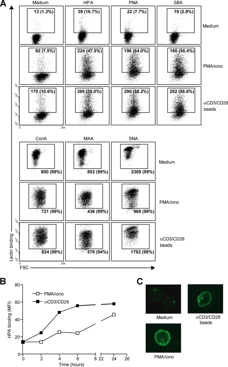
MGL ligand up-regulation is accompanied by changes in cell surface glycosylation and exposure of GalNAc-containing epitopes. CD4+ T cells were left untreated or stimulated with PMA/ionomycin or αCD3/CD28 beads. A, T cell activation is characterized by increased exposure of Thomsen-Friedenreich (TF) and Tn antigen and a decrease in overall sialylation. After overnight stimulation, binding of a well characterized panel of plant/invertebrate lectins was assessed by flow cytometry. Glycan specificities of the lectins used are as follows: HPA, α-GalNAc/Tn, PNA, Galβ1–3GalNAc, SBA, α/β-GalNAc, concanavalin A, mannose structures and di-antennary N-glycans, MAA II, α2–3-sialic acid, and SNA, α2–6-sialic acid. Mean fluorescence and the % of positive cells are indicated in the plots. Experiments were repeated six times with different donors, yielding similar results. Data are from one representative donor. FSC, forward scatter. B, Tn antigens appear 4 h after T cell stimulation with PMA/ionomycin or αCD3/CD28 beads. At each time point HPA binding was measured by flow cytometry. One independent donor out of three is shown. C, Tn antigens are localized to the surface of activated T cells. HPA staining of umstimulated as well as PMA/ionomycin or αCD3/CD28 beads activated T cells was evaluated by HPA staining and confocal microscopy.

