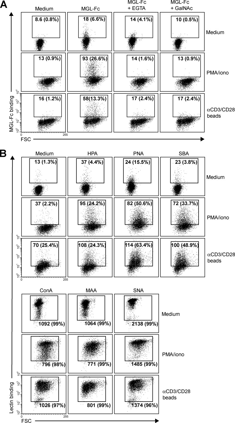
FIGURE 4.
MGL ligands are up-regulated on recently activated CD8+ T cells. CD8+ T cells were left untreated or stimulated with PMA/ionomycin or αCD3/CD28 beads. After overnight incubation, binding of MGL-Fc (A) and a panel of plant/invertebrate lectins (B) was determined by flow cytometry. Glycan specificities of the lectins used are as follows: HPA, α-GalNAc/Tn, PNA, Galβ1–3GalNAc, SBA, α/β-GalNAc, concanavalin A, mannose structures and di-antennary N-glycans, MAA II, α2–3-sialic acid and SNA, and α2–6-sialic acid. Mean fluorescence and the % of positive cells are indicated in the plots. Experiments were repeated three times with different donors, yielding similar results. One representative donor is shown. FSC, forward scatter.

