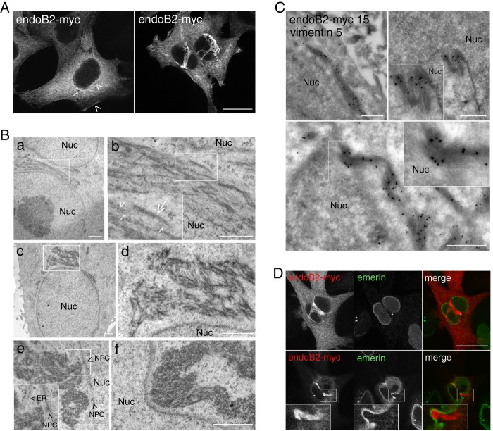
FIGURE 1.
Overexpression of endoB2 triggers the accumulation of filamentous structures around nuclei and is partly composed of intermediate filaments. HeLa cells were transfected to express endoB2 fused to a C-terminal Myc tag. A, anti-Myc immunofluorescence of z-sections with low expression level of endoB2 (left panel) showing cytosolic and discrete perinuclear as well as plasma membrane localization (arrowheads). At high expression level (right), endoB2 accumulates mainly as filamentous structures surrounding nuclei. Note that the two images were taken with different settings of the camera. z-sectioning through nuclei attested that cells are not binucleated. Scale bar, 20 μm. B, electron microscopy of Epon-embedded cells. Panels b and d correspond to magnifications of the areas delimited in (panels a and c) and show the presence of filaments of 5–10 nm diameter (arrowheads in panel b) and of 20–25 nm diameter (arrow in panel b). Panels e and f, note the proximity of bunches of filaments to nuclear membranes and nucleopore complexes (NPC (arrowheads in panel e)). ER, endoplasmic reticulum; Nuc, nucleus. Scale bars, panels a and c–f, 1 μm; panel b, 0.5 μm. C, immunoelectron microscopy using anti-Myc or anti-vimentin antibodies. Magnified areas at top right and bottom panels show the very close proximity of anti-Myc immunoreactivity to the nucleus. Nuc, nucleus; 5 nm gold, anti-vimentin; 15 nm gold, anti-Myc. Scale bar, 0.5 μm. D, anti-Myc and anti-emerin (a protein of the inner nuclear membrane) immunofluorescence of two z-sections at planes between the nucleus and the membrane contacting the coverslip (top images) or crossing the top of the nucleus (bottom images). Insets show invagination of the nuclear membrane in close contact with endoB2-myc staining. Scale bar, 20 μm.

