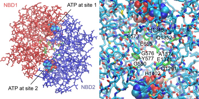
FIGURE 10.

Three-dimensional model of the CFTR NBD1-NBD2 heterodimer. The model was constructed as described under “Experimental Procedures.” Left, stick model of the overall heterodimer structure with NBD1 in red and NBD2 in blue. Two ATP molecules (in a space-filling representation) are bound between the Walker A motif of one NBD and the ABC signature motif of the other NBD. A central space between the two NBDs is evident, into which one adenosine moiety of Ap5A could extend. Residues lining this space from both NBDs are depicted in green. Right, close-up view of the central space region between the two NBDs. The residues in green lining the cavity might interact with AMP. Histidine 1348 might prevent Ap5A from interacting with ATP-binding site 1. See supplemental Movie S1 to facilitate visualizing the tree-dimensional positioning of these residues.
