Fig. 4.

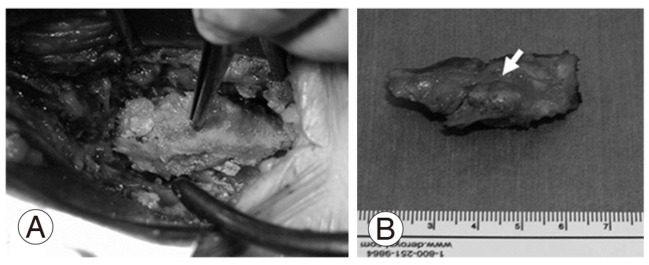
Macroscopic image of the calcified lesion excised from the ligamentum flavum. (A) Resection of the lateral recess at the L4/5 level. (B) Dorsal aspect of the lateral recess. The arrow shows a huge calcified mass in the ligamentum flavum.

Macroscopic image of the calcified lesion excised from the ligamentum flavum. (A) Resection of the lateral recess at the L4/5 level. (B) Dorsal aspect of the lateral recess. The arrow shows a huge calcified mass in the ligamentum flavum.