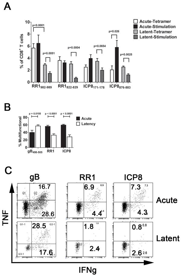
Figure 1. Subdominant CD8+ T cells become functionally compromised during latent HSV-1 infection.
TG were obtained from HSV-1 infected mice during acute (8 dpi) or latent (30–35 dpi) infection, and the dispersed cells (A) were stained with H2-Kb tetramers containing peptides corresponding to two subdominant epitopes on HSV-1 ribonucleotide reductase 1 (RR1) or infected cell protein 8 (ICP8); or were stimulated with B6WT3 fibroblasts pulsed with the same peptides, stained for intracellular IFN-γ, and analyzed by flow cytometry. The bars represent the cumulative mean ± SEM percent of CD8+ T cells that are tetramer positive during acute (2 experiments, total n = 8–10 TG per peptide) and latent (4 experiments, total n ≥ 20 TG/peptide) infection, or frequency of CD8+ T cells that are IFN-γ positive during acute or latent infection (2 experiments, n ≥ 8 TG/peptide). (B&C) TG cells were stimulated with B6WT3 fibroblasts pulsed with 3 subdominant epitopes on RR1 (RR1982-989, RR1822-829, and RR1372-380) or on ICP8 (ICP8171-178, ICP8168-176, and ICP8876-883) and monitored for surface CD107a (lytic granule release) and intracellular IFN-γ and TNFα by flow cytometry. Bars represent the cumulative mean ± SEM frequency of multifunctional (IFN-γ+, TNFα+ and CD107a+) cells (2 experiments, n ≥ 9 TG/peptide pool). (C) Representative dot plots displaying the loss of multi-functionality in RR1- and ICP-specific CD8+ T cells from acute infection to latent infection. The p values were determined by an unpaired Student’s T test.

