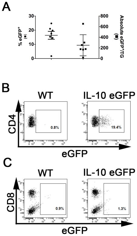
Figure 2. TG-resident CD4+ T cells produce IL-10 during latent HSV-1 infection.
TG were obtained from latently HSV-1 infected WT C57BL/6 and B6.129S6-Il10tm1Flv/Jmice, dispersed into single-cell suspensions, and stained for expression of the surface markers CD45, CD8α, CD4, and CD11c. Detection of surface marker expression and intracellular eGFP expression (from the IL-10 promoter) was performed on live cells using flow cytometry. (A) Scatter plot illustrates mean ± SEM frequency(left axis)and absolute number (right axis)of eGFP expressing CD4+CD11c− cells in individual TG (2 experiments, total n = 7). (B & C) Representative dot plots comparing the eGFP populations gated on CD4+CD11c- cells (B) or CD45+CD4- cells(c) from WT and B6.129S6-Il10tm1Flv/J.

