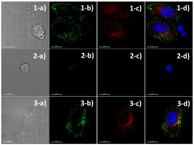
Figure 5.
Fluorescent microscopic (b-d) and contrast (a) images of HeLa/HEK mixed cells (1), HEK cells (2), and HeLa cells (3) incubated with the semi-flexible PPB/HA for 1 h, respectively. HeLa cells were fluorescently pre-labeled with a red dye (column c) before the core-shell nanoparticle incubation. Core-shell nanoparticles were seen under the green channel (column b); the nuclei were stained with a blue dye; and merged images were seen in column d. The core-shell nanoparticles preferentially labeled HeLa cells while exhibiting low binding to normal HEK cell.

