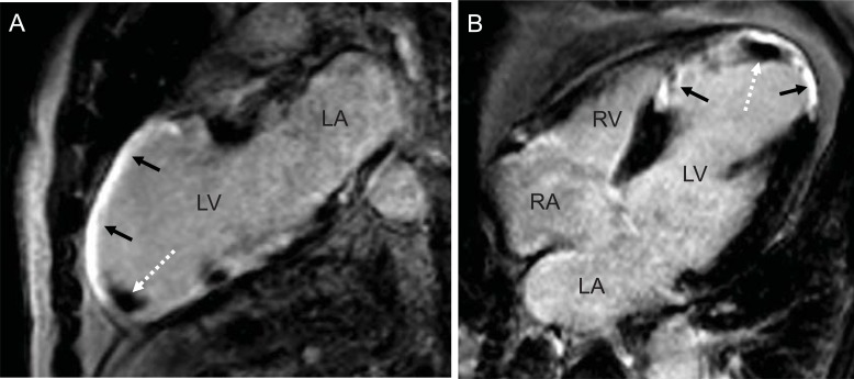
Fig. (1).
Two - (A) and four-chamber-view (B) of a patient with coronary artery disease and history of a large anterior and septal myocardial infarction (black arrows) with development of a small thrombus in the apex (white dotted arrow). (LV: left ventricle, LA: left atrium, RA: right atrium, RV: right ventricle).

