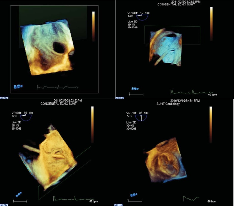
Fig. (1).
Images captured during real time three dimensional transoesophageal echocardiography for device closure of patent foramen ovale and atrial septal defects. Image in top corner left demonstrates the anatomy of an atrial septal defect from right atrial aspect with inferior vena cava entering inferiorly. Next image to the right demonstrates catheter passing through a patent foramen ovale from right atrial aspect and the following image demonstrates the catheter entering from the left atrial aspect in the same patient. Final image demonstrating device in cross section from left atrial aspect.

