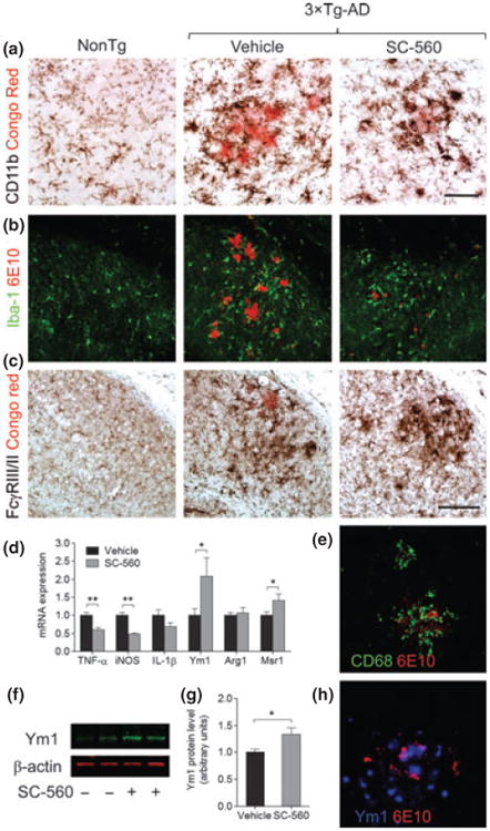
Fig. 1.

SC-560 treatment modulates microglial activation state. Representative immunostaining of microglial cells in the hippocampal subiculum of Non-Tg and 3 × Tg-AD mice treated with vehicle or SC-560. Double-staining of CD11b (a), Iba-1 (b), FcγRIII/II (c), CD68 (e), Ym1 (h), and amyloid deposits (Congo red or 6E10) showing reactive microglial cells surrounding amyloid plaques. Scale bar: a, e, h, 50 μm; b, c, 100 μm. (d) Expression of mRNA for pro-inflammatory factors and alternative activation markers in the brains of 3 × Tg-AD mice measured by quantitative real-time PCR. (f) Representative western blot of Ym1 in the brain homogenates 3 × Tg-AD mice treated with vehicle or SC-560. β-actin was used as loading control. (g) Quantification of Ym1 expression. Data shown are the means ± SEM (n = 6 mice per group). *p < 0.05 versus vehicle-treated 3 × Tg-AD mice; **p < 0.01 versus vehicle-treated 3 × Tg AD mice.
