
Figure 2.
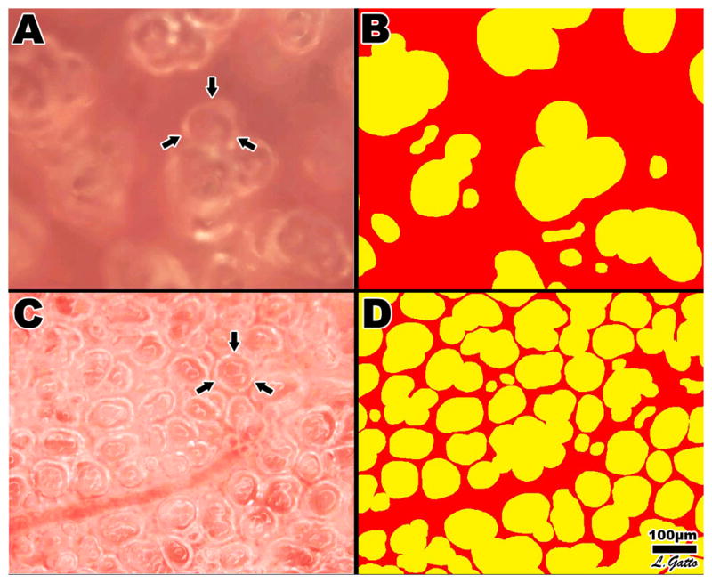
(A–D) – A: PaO2/FiO2 ratio (P/F) over time in the volume cycled ventilation (VC=▲) and airway pressure release ventilation (APRV=■) groups. There was a significant fall in P/F in the VC as compared with APRV group, with the P/F falling below 200 at T300 indicating the development of ARDS. B: Peak airway pressure (PIP) over time in the volume cycled ventilation (VC=▲) and airway pressure release ventilation (APRV=■) groups. C: Mean arterial blood pressure (MAP) over time in the volume cycled ventilation (VC=▲) and airway pressure release ventilation (APRV=■) groups. D: Airway Pressure/Time Profile (P/TP) over time in the volume cycled ventilation (VC=▲) and airway pressure release ventilation (APRV=■) groups. P/TP was significantly elevated in the APRV as compared with VC group throughout the entire experiment. BL=Baseline, EQ=Equilibrium, HS=Hemorrhagic Shock, RES=Resuscitation. Data±SEM. *=p<0.05 vs. VC group.