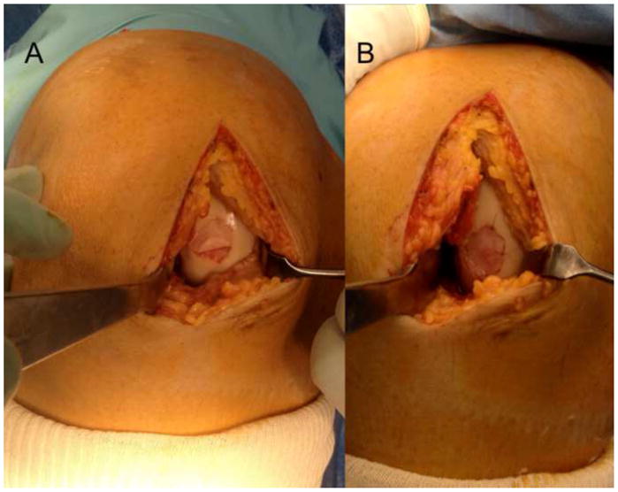
Figure 2.

After preparation of the subchondral bone and sizing of the defect, the membrane is cut to size and carefully lifted onto the defect. The membrane is glued into place using a thin layer of fibrin glue and kept in place using digital pressure. The graft should be contained inside the defect and not ride over the rim of the prepared defect. Some authors will choose to secure the edges of the graft with a 6-0 resorbable suture, however, this is not required for the technique. (Courtesy of Peter Verdonk, Antwerp, Belgium)
