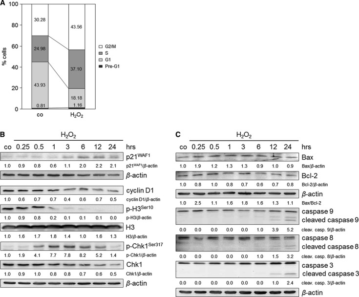
Fig. 2.

H2O2 induces cell cycle arrest and minor apoptosis in TE7 cells after treatment with 250 μM H2O2. (A) H2O2 induces S and G2 arrests 24 hrs after H2O2 treatment. Cell cycle profiles were determined by fluorescence-activated cell sorting (FACS) analysis, and the percentage of cells in the different cell cycle phases was calculated. The data are representative of three independent experiments. (B) H2O2 exposure induces an upregulation of p21WAF1, downregulation of cyclin D1 and p-H3Ser10 and activation of Chk1. Cells were cultured with H2O2 and grown for 0.25, 0.5, 1, 3, 6, 12 and 24 hrs. Lysates were immunoblotted and probed with anti-p21WAF1, -cyclin D1, -H3, -p-H3Ser10, -Chk1 and -p-Chk1Ser317 antibodies. β-actin was used as a loading control. Fold expression changes are given below the blots. (C) H2O2 initiates apoptosis pathways. Cells were incubated with H2O2 and lysates were immunoblotted for Bax, Bcl-2, caspases 8, 9 and 3 after 0.25, 0.5, 1, 3, 6, 12 and 24 hrs. The ratio of Bax/Bcl-2 is shown. β-actin was used as a loading control. Fold expression changes are given below the blots.
